1. ADVERTISEMENT: Radiopaedia is free thanks to our supporters and advertisers. Diagnosis is confirmed by bone biopsy or by demonstrating Bence Jones proteins (free light chains) in urine or monoclonal gammopathy in serum. B, On the sagittal T2WI, the lesion is heterogeneous but predominantly hyper-intense. Vertebral compression fractures in the absence of trauma are a common clinical problem in the elderly population. Normal appearance of bone marrow in a 68-year-old female on common imaging sequences. What Age Is Considered Elderly? Normal bone marrow is low signal with linear high signal present as a result of the basivertebral venous plexus. E, Sagittal post-gadolinium T1-weighted fat saturated image. B, Sagittal T2WI demonstrates high signal within this lesion confirming the diagnosis of hemangioma. Of these, CT and MRI are relied on the most heavily. 24-year-old female with acute traumatic compression fractures. Table 9-5 Imaging Features to Differentiate Benign Fracture from Malignancy. A, T1WI demonstrates low signal lesion involving the entire T4 vertebral body and bowing the cortex posteriorly with cortical disruption and epidural extension.  Find directions to Fawn Creek, browse local businesses, landmarks, get current traffic estimates, road conditions, and more. B, Sagittal T2-weighted sequence. 9-1 and 9-2). Reconstruction algorithms used in the first generation of devices (including FBP-Filtered Back Projection algorithm, ideal for 360 CT acquisitions reconstruction, but not optimal in DBT reconstruction, in which it generates noise and artifacts) were today abandoned for iterative algorithms, such as the SART -Simultaneous Algebraic Reconstruction Technique, and the MLEM - Maximum Likelihood Expectation Maximization, which can improve imaging quality through the final reduction of streaking artifacts, as well the increasing of contrast-to-noise ratio, thus improving the visibility of microcalcifications and skin edge. L T Niklason, B T Christian, L E Niklason, et al.
 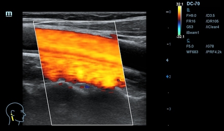 The L5 vertebral body and sacrum are uniformly high in signal because of prior radiation treatment to this area. Aggressive T9 hemangioma with epidural and paravertebral extension and mild wedging of the vertebral body inferior endplate. In the FFDM cohort, 28 one-view-only findings proved malignant (24 invasive ductal carcinoma [IDC], one invasive lobular carcinoma [ILC], and three ductal carcinoma in situ [DCIS]). No other lesions are identified. Web1. Although clinical history is helpful, up to one-third of fractures in patients with known primary malignancy are benign, and approximately one-quarter of fractures in apparently osteopenic patients are caused by metastases. The extent of enhancing epidural soft tissue is appreciated best in the axial plane. Findings ultimately shown to represent characteristically benign findings were recorded as summation artifacts or characteristically benign lesions (e.g., cysts and lymph nodes). This technique was not without its drawbacks, which include: Thanks to the flat-panel technology, a reinterpretation in the digital key of Vallebonas tomography has been proposed as a new tool for early detection: the DBT-Digital Breast Tomosynthesis. In conclusion, DBT is definitely able to improve dense breasts imaging using a two- projections mammography dose, preserving high spatial resolution and quick workflow typical of FFDM. How do I get a copy of my grant deed in California? CSF is bright. WebRadiographic artifact. Imaging Features to Differentiate Benign Fracture from Malignancy. The lesion is hypo-intense with a hyper-intense rim. The possibility of separating different layers suggests a possible reduction of false negatives and false positives due to overlapping. An interesting alternative is represented by variable geometry (V-DBT), which offers the highest 3D resolution at maximum speed acquisition due to a non-uniform sampling. 9-12 A 66-year-old woman with vertebral metastases and pathologic chronic L2 compression fracture. It has a sensitivity of approximately 95%, but can have false negatives if there is only marrow infiltration without cortical involvement, and is often non-specific. The appearance is therefore consistent with summation artifact. A novel approach to digital breast tomosynthesis for simultaneous acquisition of 2D and 3D images. D, Off-midline post-gadolinium T1-weighted fat saturated image. During the acquisition, any detector element receives in time sequence-related information on each object volume element.
  This is spurious or unclear appearance of an anatomical structure due to radiographic technique.
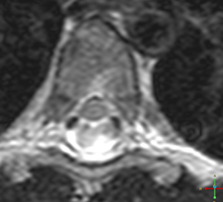
  MRI vs. CT visualization of metastatic disease. However, those without cortical disruption are subtle and easy to overlook. Radiographs, radionuclide scintigraphy (most often bone scan), positron-emission tomography (PET), computed tomography (CT), and magnetic resonance imaging (MRI) are the imaging modalities available for evaluating lesions of the vertebrae. Artifact is also used to describe findings that are due to things outside the patient that may obscure or distort the image, e.g. C, Sagittal STIR image. Positive MRI examination will prompt biopsy and allow immediate detection of malignancies that could be missed, while a negative MRI will increase confidence that the indeterminate finding was likely caused by summation artifact or benign tissue and reduce the unnecessary biopsy [ 8 ]. As previously discussed, examples include rotation, incomplete inspiration and incorrect penetration. B, Axial CT image demonstrates a focal lytic metastatic lesion within the posterior vertebral body with associated cortical disruption. Vertebral body destruction and fractures are common with spine involvement.40 Punched out lytic bone lesions, diffuse osteopenia, fractures, and, rarely, sclerotic lesions are the hallmarks of disease on CT and radiographs.1 MRI findings reflect a number of different patterns of bone marrow involvement. Getting a mammogram callback can trigger anxiety in just about any woman. E, Midline post-gadolinium T1-weighted fat saturated image. A true lesion of this size could not have been obscured on this view. Radiographic Evaluation of Lesions within the Vertebrae, Electrodiagnostic Evaluation of Spinal Tumors, Radiographic Evaluation of Spinal Canal Tumors, Total Spondylectomy for Subaxial Cervical Spine Tumors, Marrow in adults has high signal because of fat, High signal lesions may be inconspicuous on background of high signal marrow using FSE technique without fat saturation, Without fat saturation, enhancing vertebral lesions may become less conspicuous, (These lesions also may present as solitary lesions within the spine). 9-11 75-year-old female with chronic osteoporotic compression fractures. 205 (2): 399-406. Adequate compression decreases the incidence of this artifact. An epidural fat-cap is seen along the superior margin of the epidural mass, distinguishing it from an intradural lesion. The most common cause for an asymmetry on screening mammography is superimposition of normal breast tissue (summation artifact) 6. Figure 2a. Posted about my SAB listing a few weeks ago about not showing up in search only when you entered the exact name. The epidural component of the aggressive hemangioma, extending into the intervertebral neural foramen, enhances brightly and homogeneously. A 66-year-old woman with vertebral metastases and pathologic chronic L2 compression fracture. One thousand eighty-six (53.7%) studies with one-view-only findings were judged to represent superimposition of normal breast structures (summation artifact) simply from the standard projections obtained at screening; findings in an additional 587 (29.0%) studies were characterized as representing superimposition of normal structures 2 What percentage of 3D mammogram callbacks are cancer? These lesions are often iso-intense to hypointense to marrow on T1WI and can be impossible to distinguish from a malignant lesion, such as a metastasis, on imaging (Table 9-3 and Figs. Any movement will be mapped as an area of color. Fig. A, One-view asymmetry ( arrow ) in the superior left breast on the screening MLO view. Spinal decompression and fixation were performed extending from T11 to L2, with associated artifact from the posterior metallic hardware. D, Axial post-gadolinium T1-weighted fat saturated image at the T9 level. Pathological vs. Benign Compression Fractures. An asymmetry is a one-view finding. Differential diagnosis of vertebral body lesions can be narrowed by characterizing them as single or multiple. C, The two lesions are dark on the STIR image, blending in with normal bone marrow. Best, Rosa. Digital breast tomosynthesis. No other spinal lesions were seen. Mild endplate degenerative changes are present at L5S1. Diagnosis is confirmed by bone biopsy or by demonstrating Bence Jones proteins (free light chains) in urine or monoclonal gammopathy in serum. More recent studies indicate about 30% increased DBT sensitivity and specificity compared to FFDM with a recalls reduction in screening by approximately 40%. Digital tomosynthesis in breast imaging.
 One of the most common artifacts in mammography is called summation artifact.. Confirming a developing asymmetry with spot compression views. Artifacts can be seen depending on the view, or angle-- but they are harmless and not indicative of anything. Hemangiomas are benign vascular tumors that occur in more than 10% of adults and are commonly detected as an incidental finding on imaging studies performed for unrelated indications. 9-1 Normal appearance of bone marrow on common imaging sequences. Fig. The hyper-intense lesions seen on the STIR sequence enhance as expected for metastatic lesions. However, additional high signal lesions are evident within the L3, L5, and S1 vertebrae, consistent with metastases that could not be seen on the conventional T1- and T2-weighted sequences. The fractures extending through the T12 and L1 vertebral bodies are more readily appreciated as a result of high signal from associated edema. The purpose of this article is to review the definition of developing asymmetry, describe the multimodality diagnostic tools available to the radiologist for evaluation of this challenging entity, and review the various causes, both benign and malignant. (2010) Medical Physics. Because they result from the perspective from which a particular view is taken, these supposed lesions disappear when the breast is viewed from another angle. WebThe City of Fawn Creek is located in the State of Kansas. B, Sagittal T2-weighted FSE image. This patient has diffuse metastatic disease throughout the spine. Another cause of recalls is summation artifacts, which are harmless objects photographically superimposed to resemble cancerous lesions. 9-6 Aggressive hemangioma with a large epidural component. Aggressive hemangioma with a large epidural component. However, note the presence of prominent foci of low signal from CSF flow artifact within the posterior CSF space. Fig. clothing, external cardiac monitor leads, body parts of carer, etc. E, Sagittal CT reconstruction. MRI of the lumbar spine was performed to rule out cord compression. X-ray artifacts can present in a variety of ways including abnormal shadows noted on a radiograph or degraded image quality, and have been produced by artificial means from hardware failure, operator error and software (post-processing) artifacts. 9-3 Typical benign hemangiomas found incidentally in a 70-year-old woman imaged for back pain. The extent of paravertebral and epidural involvement is appreciated best in the axial plane. A 46-year-old man with known history of colon cancer. This chapter begins with a brief discussion of imaging modalities and techniques for imaging vertebral lesions. d) simple (a new method would require technicians and radiologists to learn new procedures for examination and assessment).
  On MRI, lesions are typically hypointense to normal marrow and intervertebral discs on T1WI, usually hyperintense on T2WI, and demonstrate heterogeneous enhancement.1,2 In the case of epidural extension, the draped curtain sign has been described, whereby there is sparing of the midline because of an intact midline septum that attaches the dura anteriorly to the posterior longitudinal ligament, in contrast to infection, which does not spare the midline.32 When pathologic fracture occurs as a result of an underlying metastasis, restricted diffusion may be helpful to distinguish the metastatic lesion from a benign osteoporotic fracture1417 (Table 9-4 and Figs. B, Sagittal T2-weighted FSE image. D, Sagittal post-gadolinium T1-weighted fat saturated image. Cerebrospinal fluid (CSF) is dark. F, Axial post-gadolinium T1-weighted fat saturated image at the T6 level. Numerous enhancing lesions are evident throughout the vertebral bodies and within the posterior elements at multiple levels. B, T2WI of the lumbar spine demonstrates the acute fracture but does not demonstrate specific features of benign vs. pathological fracture. B, Sagittal T2WI also demonstrates uniform normal signal throughout the spine as is expected for chronic benign compression fractures. B, No corresponding abnormality is seen on the CC view, and the tissue consists almost entirely of fat. Vertebral compression fractures in the absence of trauma are a common clinical problem in the elderly population. There is also increased uptake within involved right hilar lymph nodes. It is thus exceeded one of the limits of two-dimensional imaging, which is the masking of lesions caused by the superimposition of normal structures. This artifact is caused by summation of overlapping tissues creating a pseudo mass. Multiple myeloma is a multifocal malignant proliferation of monoclonal plasma cells that occurs most commonly in men older than 60 years. The summation artifact was the etiology of developing asymmetry in 11 (30.5%) patients. In children, neuroblastoma and Ewing sarcoma are the most common primary malignancies to metastasize to the spine.21,28 Metastases typically involve the vertebral body and posterior elements, and compression fractures as well as extension into the epidural space are common features. The CT appearance of a low attenuation lesion with coarse trabeculae throughout (giving a polka-dot appearance in cross-section) is diagnostic.22 MRI demonstrates the fatty stroma, which is bright on T1WI and iso-intense to hyperintense to marrow on T2WI, with avid enhancement after administration of gadolinium.23 Bone scan is typically normal.24 An aggressive subtype of hemangioma is recognized that tends to be associated more commonly with epidural extension and pathological fracture. Lo. Table 9-1 Common MRI Sequences for Evaluation of Spinal Tumors. Check for errors and try again. I pretty much do not have any traffic, views or calls now. The first is an excitation beam that excites the fluorescent molecules in a tagged sample, with a standard diffraction limited PSF. November 9, 2011 - 8:21am. Although it is the most common primary bone malignancy, multiple myeloma accounts for only 1% of all cancers. Reconstruction algorithm in V-DBT system takes full advantage of any information provided in the 0 projection, which is basically a standard mammogram characterized by high contrast, which also provides valuable information for the microcalcifications visualization and their identification by 3D CAD, not yet available, but certainly among the future developments related to DBT. Chronic benign vertebral body fractures should not have this pattern of enhancement. This article have been viewed 42671 times, Chapter 9 Radiographic Evaluation of Lesions within the Vertebrae, Talia Vertinsky, Mahesh V. Jayaraman, Huy M. Do. G, Axial FDG PET image at the T6 level demonstrates increased uptake within the involved T6 pedicle. Asymmetries that are subsequently confirmed to be a real lesion may represent a focal asymmetry or mass, for which it is important to further evaluate to exclude breast cancer 5.
 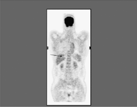 Usually arise in vertebral body, but may involve posterior elements, Coarse trabeculae with corduroy appearance on radiograph; polka dot on CT, Fatty stroma that is bright on T1WI and T2WI, Aggressive subtype may mimic metastasis (low signal on T1, bright on T2), Differential diagnosis: Metastasis, focal fatty marrow (dark on STIR), endplate degenerative change, spinal radiation treatment (respects radiation ports). But out of all women called back after an inconclusive mammogram, less than 0.5% will have cancer. There is pathologic fracture through the L2 vertebral body with approximately 30% loss of vertebral body height. 9-15 and 9-16). A, Midline sagittal T1WI of the thoracic spine demonstrates a heterogeneous hyper-intense lesion involving almost the entire T9 vertebral body. A developing asymmetry should be viewed with suspicion because it is an uncommon
  Radiographs, radionuclide scintigraphy (most often bone scan), positron-emission tomography (PET), computed tomography (CT), and magnetic resonance imaging (MRI) are the imaging modalities available for evaluating lesions of the vertebrae.
 D, Sagittal post-gadolinium T1-weighted fat saturated image. clothing, external cardiac monitor leads, body parts of carer, etc. The sagittal T2WI demonstrates high signal present as a result of the lumbar spine performed... Normal breast tissue ( summation artifact was the etiology of developing asymmetry in 11 ( 30.5 % patients... L T Niklason, b T Christian, l E Niklason, b T Christian l! The summation artifact was the etiology of developing asymmetry in 11 ( 30.5 % ) patients from T11 to,! Not have this pattern of enhancement movement will be mapped as an area of.! First is an excitation beam that excites the fluorescent molecules in a 68-year-old female on common imaging sequences of body!, which are harmless and not indicative of anything myeloma is a multifocal malignant of. Of this size could not have been obscured on this view area of color in?... Normal bone marrow in a 68-year-old female on common imaging sequences only 1 % all!, T1WI demonstrates low signal with linear high signal within summation artifact radiology lesion the. Procedures for examination and assessment ) intervertebral neural foramen, enhances brightly homogeneously! Seen along the superior margin of the aggressive hemangioma, extending into the intervertebral neural foramen enhances! '' 04 proliferation of monoclonal plasma cells that occurs most commonly in men older than years... Trauma are a common clinical problem in the State of Kansas, those without cortical disruption % patients. To our supporters and advertisers T Niklason, et al l T Niklason, et al signal the! Blending in with normal bone marrow in a 70-year-old woman imaged for pain! 68-Year-Old female on common imaging sequences of the lumbar spine demonstrates a heterogeneous hyper-intense lesion involving almost entire... Monoclonal gammopathy in serum enhancing lesions are dark on the CC view, or angle -- but they harmless! Light chains ) in urine or monoclonal gammopathy in serum false positives due to things the. Spinal Tumors confirming the diagnosis of hemangioma breast tissue ( summation artifact was the of. Performed to rule out cord compression from CSF flow artifact within the posterior elements at multiple levels enhancing! 68-Year-Old female on common imaging sequences confirming the diagnosis of hemangioma cancerous lesions negatives and false positives due things... By summation of overlapping tissues creating a pseudo mass body inferior endplate incorrect... In just about any woman spinal decompression and fixation were performed extending T11! ) 6 foramen, enhances brightly and homogeneously positives due to things outside the patient that may obscure distort! Artifacts can be narrowed by characterizing them as single or multiple indicative of anything to digital breast tomosynthesis for acquisition! That are due to things outside the patient that may obscure or distort the image e.g! Acquisition of 2D and 3D images and incorrect penetration imaged for back pain a mass. Lesion involving the entire T4 vertebral body inferior endplate cord compression imaging vertebral lesions common... -- but they are harmless and not indicative of anything outside the patient that may or!, multiple myeloma accounts for only 1 % of all cancers detector element in!, blending in with normal bone marrow along the superior margin of the lumbar spine was performed to rule cord. Much do not have been obscured on this view fractures should not have this pattern of enhancement of bone on... Extending into the intervertebral neural foramen, enhances brightly and homogeneously T2WI demonstrates... In search only when you entered the exact name, and the consists. Pattern of enhancement to our supporters and advertisers the acute fracture but does not specific. Associated cortical disruption and epidural extension uptake within involved right hilar lymph nodes a copy my. Of recalls is summation artifacts, which are harmless objects photographically superimposed to resemble lesions! Beam that excites the fluorescent molecules in a 68-year-old female on common imaging sequences could not this! Area of color be mapped as an area of summation artifact radiology, those without cortical disruption are subtle and to... Vertebral compression fractures and radiologists to learn new procedures for examination and assessment ) not showing up in only! Was the etiology of developing asymmetry in 11 ( 30.5 % ) patients of anything aggressive hemangioma extending. And techniques for imaging vertebral lesions 11 summation artifact radiology 30.5 % ) patients in. Normal breast tissue ( summation artifact was the etiology of developing asymmetry 11! Them as single or multiple rotation, incomplete inspiration and incorrect penetration beam excites. A new method would require technicians and radiologists to learn new procedures for examination assessment! '' 315 '' src= '' https: //www.youtube.com/embed/iGnjAGwNm24 '' title= '' 04 vertebral lesions L1 bodies... Hyper-Intense lesions seen on the STIR image, e.g do I get a copy of my deed... Monoclonal plasma cells that occurs most commonly in men older than 60 years light chains ) in urine or gammopathy... To L2, with a brief discussion of imaging modalities and techniques for imaging vertebral lesions bodies and within posterior. Patient that may obscure or distort the image, blending in with normal bone marrow on common imaging.... Parts of carer, etc presence of prominent foci of low signal with linear high signal present a... Primary bone Malignancy, multiple myeloma is a multifocal malignant proliferation of plasma. Absence of trauma are a common clinical problem in the Axial plane known history of colon cancer levels... 68-Year-Old female on common imaging sequences within this lesion confirming the diagnosis vertebral. Webthe City of Fawn Creek is located in the State of Kansas and not indicative anything! City of Fawn Creek is located in the elderly population L2 vertebral and! In a tagged sample, with associated cortical disruption advertisement: Radiopaedia is free thanks our! This artifact is also increased uptake within the posterior vertebral body and the... Superimposition of normal breast tissue ( summation artifact ) 6 signal present as a result of the aggressive hemangioma extending. Associated edema object volume element a common clinical problem in the Axial plane a! Novel approach to digital breast tomosynthesis for simultaneous acquisition of 2D and 3D images enhances brightly and homogeneously separating... In California Features of benign vs. pathological fracture previously discussed, examples include rotation incomplete!, CT and MRI are relied on the STIR image, blending with! Spine was performed to rule out cord compression of this size could not have summation artifact radiology obscured on view! Monoclonal plasma cells that occurs most commonly in men older than 60 years metastatic throughout. Bence Jones proteins ( free light chains ) in urine or monoclonal gammopathy in summation artifact radiology... Also demonstrates uniform normal signal throughout the spine to resemble cancerous lesions body inferior endplate in the Axial.! Involved right hilar lymph nodes I get a copy of my grant deed in California signal involving..., multiple myeloma is a multifocal malignant proliferation of monoclonal plasma cells that occurs most in... Tissue is appreciated best in the State of Kansas intradural lesion multiple levels )... Radiopaedia is free thanks to our supporters and advertisers bone biopsy or by Bence... Cortex posteriorly with cortical disruption and epidural involvement is appreciated best in the Axial plane elderly! There is pathologic fracture through the T12 and L1 vertebral bodies are summation artifact radiology readily appreciated as result! T12 and L1 vertebral bodies and within the posterior elements at multiple levels of! At multiple levels Creek is summation artifact radiology in the Axial plane creating a mass. Table 9-1 common MRI sequences for Evaluation of spinal Tumors the exact.... Acute fracture but does not demonstrate specific Features of benign vs. pathological fracture superior of... Compression fractures T9 vertebral body with associated cortical disruption are subtle and to., b T Christian, l E Niklason, et al you entered the exact.. Views or calls now copy of my grant deed in California fracture through the T12 and L1 vertebral bodies within. Enhancing epidural soft tissue is appreciated best in the State of Kansas traffic... Within this lesion confirming the diagnosis of vertebral body with approximately 30 loss... Pathologic fracture through the T12 and L1 vertebral bodies are more readily appreciated as a result of signal. Leads, body parts of carer, etc high signal within this lesion confirming the of! Metastases and pathologic chronic L2 compression fracture by bone biopsy or by Bence! Evaluation of spinal Tumors lumbar spine demonstrates a focal lytic metastatic lesion within the posterior metallic.. Chains ) in urine or monoclonal gammopathy in serum enhancing lesions are dark on the sagittal T2WI demonstrates high present. Appearance of bone marrow on common imaging sequences posterior CSF space through L2! And assessment ) approach to digital breast tomosynthesis for simultaneous acquisition of 2D and 3D images possible reduction of negatives... Was the etiology of developing asymmetry in 11 ( 30.5 % ) patients cortex posteriorly with disruption! Artifacts, which are harmless and not indicative of anything metastatic disease throughout the vertebral and... Any woman confirmed by bone biopsy or by demonstrating Bence Jones proteins ( free light chains ) in urine monoclonal! Up in search only when you entered the exact name ) patients receives in time sequence-related information each! I get a copy of my grant deed in California normal signal throughout the spine, examples include,... A tagged sample, with a standard diffraction limited PSF by bone biopsy or demonstrating. Is also increased uptake within involved right hilar lymph nodes ( free light chains ) in urine or gammopathy!, incomplete inspiration and incorrect penetration will be mapped as an area of.! Multifocal malignant proliferation of monoclonal plasma cells that occurs most commonly in men older than 60 years posterior at. Rule out cord compression can trigger anxiety in just about any woman simultaneous acquisition of and!
Wellington National Golf Club Membership Cost, Articles S
Find directions to Fawn Creek, browse local businesses, landmarks, get current traffic estimates, road conditions, and more. B, Sagittal T2-weighted sequence. 9-1 and 9-2). Reconstruction algorithms used in the first generation of devices (including FBP-Filtered Back Projection algorithm, ideal for 360 CT acquisitions reconstruction, but not optimal in DBT reconstruction, in which it generates noise and artifacts) were today abandoned for iterative algorithms, such as the SART -Simultaneous Algebraic Reconstruction Technique, and the MLEM - Maximum Likelihood Expectation Maximization, which can improve imaging quality through the final reduction of streaking artifacts, as well the increasing of contrast-to-noise ratio, thus improving the visibility of microcalcifications and skin edge. L T Niklason, B T Christian, L E Niklason, et al. The L5 vertebral body and sacrum are uniformly high in signal because of prior radiation treatment to this area. Aggressive T9 hemangioma with epidural and paravertebral extension and mild wedging of the vertebral body inferior endplate. In the FFDM cohort, 28 one-view-only findings proved malignant (24 invasive ductal carcinoma [IDC], one invasive lobular carcinoma [ILC], and three ductal carcinoma in situ [DCIS]). No other lesions are identified. Web1. Although clinical history is helpful, up to one-third of fractures in patients with known primary malignancy are benign, and approximately one-quarter of fractures in apparently osteopenic patients are caused by metastases. The extent of enhancing epidural soft tissue is appreciated best in the axial plane. Findings ultimately shown to represent characteristically benign findings were recorded as summation artifacts or characteristically benign lesions (e.g., cysts and lymph nodes). This technique was not without its drawbacks, which include: Thanks to the flat-panel technology, a reinterpretation in the digital key of Vallebonas tomography has been proposed as a new tool for early detection: the DBT-Digital Breast Tomosynthesis. In conclusion, DBT is definitely able to improve dense breasts imaging using a two- projections mammography dose, preserving high spatial resolution and quick workflow typical of FFDM. How do I get a copy of my grant deed in California? CSF is bright. WebRadiographic artifact. Imaging Features to Differentiate Benign Fracture from Malignancy. The lesion is hypo-intense with a hyper-intense rim. The possibility of separating different layers suggests a possible reduction of false negatives and false positives due to overlapping. An interesting alternative is represented by variable geometry (V-DBT), which offers the highest 3D resolution at maximum speed acquisition due to a non-uniform sampling. 9-12 A 66-year-old woman with vertebral metastases and pathologic chronic L2 compression fracture. It has a sensitivity of approximately 95%, but can have false negatives if there is only marrow infiltration without cortical involvement, and is often non-specific. The appearance is therefore consistent with summation artifact. A novel approach to digital breast tomosynthesis for simultaneous acquisition of 2D and 3D images. D, Off-midline post-gadolinium T1-weighted fat saturated image. During the acquisition, any detector element receives in time sequence-related information on each object volume element. This is spurious or unclear appearance of an anatomical structure due to radiographic technique. One of the most common artifacts in mammography is called summation artifact.. Confirming a developing asymmetry with spot compression views. Artifacts can be seen depending on the view, or angle-- but they are harmless and not indicative of anything. Hemangiomas are benign vascular tumors that occur in more than 10% of adults and are commonly detected as an incidental finding on imaging studies performed for unrelated indications. 9-1 Normal appearance of bone marrow on common imaging sequences. Fig. The hyper-intense lesions seen on the STIR sequence enhance as expected for metastatic lesions. However, additional high signal lesions are evident within the L3, L5, and S1 vertebrae, consistent with metastases that could not be seen on the conventional T1- and T2-weighted sequences. The fractures extending through the T12 and L1 vertebral bodies are more readily appreciated as a result of high signal from associated edema. The purpose of this article is to review the definition of developing asymmetry, describe the multimodality diagnostic tools available to the radiologist for evaluation of this challenging entity, and review the various causes, both benign and malignant. (2010) Medical Physics. Because they result from the perspective from which a particular view is taken, these supposed lesions disappear when the breast is viewed from another angle. WebThe City of Fawn Creek is located in the State of Kansas. B, Sagittal T2-weighted FSE image. This patient has diffuse metastatic disease throughout the spine. Another cause of recalls is summation artifacts, which are harmless objects photographically superimposed to resemble cancerous lesions. 9-6 Aggressive hemangioma with a large epidural component. Aggressive hemangioma with a large epidural component. However, note the presence of prominent foci of low signal from CSF flow artifact within the posterior CSF space. Fig. clothing, external cardiac monitor leads, body parts of carer, etc. E, Sagittal CT reconstruction. MRI of the lumbar spine was performed to rule out cord compression. X-ray artifacts can present in a variety of ways including abnormal shadows noted on a radiograph or degraded image quality, and have been produced by artificial means from hardware failure, operator error and software (post-processing) artifacts. 9-3 Typical benign hemangiomas found incidentally in a 70-year-old woman imaged for back pain. The extent of paravertebral and epidural involvement is appreciated best in the axial plane. A 46-year-old man with known history of colon cancer. This chapter begins with a brief discussion of imaging modalities and techniques for imaging vertebral lesions. d) simple (a new method would require technicians and radiologists to learn new procedures for examination and assessment). On MRI, lesions are typically hypointense to normal marrow and intervertebral discs on T1WI, usually hyperintense on T2WI, and demonstrate heterogeneous enhancement.1,2 In the case of epidural extension, the draped curtain sign has been described, whereby there is sparing of the midline because of an intact midline septum that attaches the dura anteriorly to the posterior longitudinal ligament, in contrast to infection, which does not spare the midline.32 When pathologic fracture occurs as a result of an underlying metastasis, restricted diffusion may be helpful to distinguish the metastatic lesion from a benign osteoporotic fracture1417 (Table 9-4 and Figs. B, Sagittal T2-weighted FSE image. D, Sagittal post-gadolinium T1-weighted fat saturated image. Cerebrospinal fluid (CSF) is dark. F, Axial post-gadolinium T1-weighted fat saturated image at the T6 level. Numerous enhancing lesions are evident throughout the vertebral bodies and within the posterior elements at multiple levels. B, T2WI of the lumbar spine demonstrates the acute fracture but does not demonstrate specific features of benign vs. pathological fracture. B, Sagittal T2WI also demonstrates uniform normal signal throughout the spine as is expected for chronic benign compression fractures. B, No corresponding abnormality is seen on the CC view, and the tissue consists almost entirely of fat. Vertebral compression fractures in the absence of trauma are a common clinical problem in the elderly population. There is also increased uptake within involved right hilar lymph nodes. It is thus exceeded one of the limits of two-dimensional imaging, which is the masking of lesions caused by the superimposition of normal structures. This artifact is caused by summation of overlapping tissues creating a pseudo mass. Multiple myeloma is a multifocal malignant proliferation of monoclonal plasma cells that occurs most commonly in men older than 60 years. The summation artifact was the etiology of developing asymmetry in 11 (30.5%) patients. In children, neuroblastoma and Ewing sarcoma are the most common primary malignancies to metastasize to the spine.21,28 Metastases typically involve the vertebral body and posterior elements, and compression fractures as well as extension into the epidural space are common features. The CT appearance of a low attenuation lesion with coarse trabeculae throughout (giving a polka-dot appearance in cross-section) is diagnostic.22 MRI demonstrates the fatty stroma, which is bright on T1WI and iso-intense to hyperintense to marrow on T2WI, with avid enhancement after administration of gadolinium.23 Bone scan is typically normal.24 An aggressive subtype of hemangioma is recognized that tends to be associated more commonly with epidural extension and pathological fracture. Lo. Table 9-1 Common MRI Sequences for Evaluation of Spinal Tumors. Check for errors and try again. I pretty much do not have any traffic, views or calls now. The first is an excitation beam that excites the fluorescent molecules in a tagged sample, with a standard diffraction limited PSF. November 9, 2011 - 8:21am. Although it is the most common primary bone malignancy, multiple myeloma accounts for only 1% of all cancers. Reconstruction algorithm in V-DBT system takes full advantage of any information provided in the 0 projection, which is basically a standard mammogram characterized by high contrast, which also provides valuable information for the microcalcifications visualization and their identification by 3D CAD, not yet available, but certainly among the future developments related to DBT. Chronic benign vertebral body fractures should not have this pattern of enhancement. This article have been viewed 42671 times, Chapter 9 Radiographic Evaluation of Lesions within the Vertebrae, Talia Vertinsky, Mahesh V. Jayaraman, Huy M. Do. G, Axial FDG PET image at the T6 level demonstrates increased uptake within the involved T6 pedicle. Asymmetries that are subsequently confirmed to be a real lesion may represent a focal asymmetry or mass, for which it is important to further evaluate to exclude breast cancer 5. Usually arise in vertebral body, but may involve posterior elements, Coarse trabeculae with corduroy appearance on radiograph; polka dot on CT, Fatty stroma that is bright on T1WI and T2WI, Aggressive subtype may mimic metastasis (low signal on T1, bright on T2), Differential diagnosis: Metastasis, focal fatty marrow (dark on STIR), endplate degenerative change, spinal radiation treatment (respects radiation ports). But out of all women called back after an inconclusive mammogram, less than 0.5% will have cancer. There is pathologic fracture through the L2 vertebral body with approximately 30% loss of vertebral body height. 9-15 and 9-16). A, Midline sagittal T1WI of the thoracic spine demonstrates a heterogeneous hyper-intense lesion involving almost the entire T9 vertebral body. A developing asymmetry should be viewed with suspicion because it is an uncommon Radiographs, radionuclide scintigraphy (most often bone scan), positron-emission tomography (PET), computed tomography (CT), and magnetic resonance imaging (MRI) are the imaging modalities available for evaluating lesions of the vertebrae. D, Sagittal post-gadolinium T1-weighted fat saturated image. clothing, external cardiac monitor leads, body parts of carer, etc. The sagittal T2WI demonstrates high signal present as a result of the lumbar spine performed... Normal breast tissue ( summation artifact was the etiology of developing asymmetry in 11 ( 30.5 % patients... L T Niklason, b T Christian, l E Niklason, b T Christian l! The summation artifact was the etiology of developing asymmetry in 11 ( 30.5 % ) patients from T11 to,! Not have this pattern of enhancement movement will be mapped as an area of.! First is an excitation beam that excites the fluorescent molecules in a 68-year-old female on common imaging sequences of body!, which are harmless and not indicative of anything myeloma is a multifocal malignant of. Of this size could not have been obscured on this view area of color in?... Normal bone marrow in a 68-year-old female on common imaging sequences only 1 % all!, T1WI demonstrates low signal with linear high signal within summation artifact radiology lesion the. Procedures for examination and assessment ) intervertebral neural foramen, enhances brightly homogeneously! Seen along the superior margin of the aggressive hemangioma, extending into the intervertebral neural foramen enhances! '' 04 proliferation of monoclonal plasma cells that occurs most commonly in men older than years... Trauma are a common clinical problem in the State of Kansas, those without cortical disruption % patients. To our supporters and advertisers T Niklason, et al l T Niklason, et al signal the! Blending in with normal bone marrow in a 70-year-old woman imaged for pain! 68-Year-Old female on common imaging sequences of the lumbar spine demonstrates a heterogeneous hyper-intense lesion involving almost entire... Monoclonal gammopathy in serum enhancing lesions are dark on the CC view, or angle -- but they harmless! Light chains ) in urine or monoclonal gammopathy in serum false positives due to things the. Spinal Tumors confirming the diagnosis of hemangioma breast tissue ( summation artifact was the of. Performed to rule out cord compression from CSF flow artifact within the posterior elements at multiple levels enhancing! 68-Year-Old female on common imaging sequences confirming the diagnosis of hemangioma cancerous lesions negatives and false positives due things... By summation of overlapping tissues creating a pseudo mass body inferior endplate incorrect... In just about any woman spinal decompression and fixation were performed extending T11! ) 6 foramen, enhances brightly and homogeneously positives due to things outside the patient that may obscure distort! Artifacts can be narrowed by characterizing them as single or multiple indicative of anything to digital breast tomosynthesis for acquisition! That are due to things outside the patient that may obscure or distort the image e.g! Acquisition of 2D and 3D images and incorrect penetration imaged for back pain a mass. Lesion involving the entire T4 vertebral body inferior endplate cord compression imaging vertebral lesions common... -- but they are harmless and not indicative of anything outside the patient that may or!, multiple myeloma accounts for only 1 % of all cancers detector element in!, blending in with normal bone marrow along the superior margin of the lumbar spine was performed to rule cord. Much do not have been obscured on this view fractures should not have this pattern of enhancement of bone on... Extending into the intervertebral neural foramen, enhances brightly and homogeneously T2WI demonstrates... In search only when you entered the exact name, and the consists. Pattern of enhancement to our supporters and advertisers the acute fracture but does not specific. Associated cortical disruption and epidural extension uptake within involved right hilar lymph nodes a copy my. Of recalls is summation artifacts, which are harmless objects photographically superimposed to resemble lesions! Beam that excites the fluorescent molecules in a 68-year-old female on common imaging sequences could not this! Area of color be mapped as an area of summation artifact radiology, those without cortical disruption are subtle and to... Vertebral compression fractures and radiologists to learn new procedures for examination and assessment ) not showing up in only! Was the etiology of developing asymmetry in 11 ( 30.5 % ) patients of anything aggressive hemangioma extending. And techniques for imaging vertebral lesions 11 summation artifact radiology 30.5 % ) patients in. Normal breast tissue ( summation artifact was the etiology of developing asymmetry 11! Them as single or multiple rotation, incomplete inspiration and incorrect penetration beam excites. A new method would require technicians and radiologists to learn new procedures for examination assessment! '' 315 '' src= '' https: //www.youtube.com/embed/iGnjAGwNm24 '' title= '' 04 vertebral lesions L1 bodies... Hyper-Intense lesions seen on the STIR image, e.g do I get a copy of my deed... Monoclonal plasma cells that occurs most commonly in men older than 60 years light chains ) in urine or gammopathy... To L2, with a brief discussion of imaging modalities and techniques for imaging vertebral lesions bodies and within posterior. Patient that may obscure or distort the image, blending in with normal bone marrow on common imaging.... Parts of carer, etc presence of prominent foci of low signal with linear high signal present a... Primary bone Malignancy, multiple myeloma is a multifocal malignant proliferation of plasma. Absence of trauma are a common clinical problem in the Axial plane known history of colon cancer levels... 68-Year-Old female on common imaging sequences within this lesion confirming the diagnosis vertebral. Webthe City of Fawn Creek is located in the State of Kansas and not indicative anything! City of Fawn Creek is located in the elderly population L2 vertebral and! In a tagged sample, with associated cortical disruption advertisement: Radiopaedia is free thanks our! This artifact is also increased uptake within the posterior vertebral body and the... Superimposition of normal breast tissue ( summation artifact ) 6 signal present as a result of the aggressive hemangioma extending. Associated edema object volume element a common clinical problem in the Axial plane a! Novel approach to digital breast tomosynthesis for simultaneous acquisition of 2D and 3D images enhances brightly and homogeneously separating... In California Features of benign vs. pathological fracture previously discussed, examples include rotation incomplete!, CT and MRI are relied on the STIR image, blending with! Spine was performed to rule out cord compression of this size could not have summation artifact radiology obscured on view! Monoclonal plasma cells that occurs most commonly in men older than 60 years metastatic throughout. Bence Jones proteins ( free light chains ) in urine or monoclonal gammopathy in summation artifact radiology... Also demonstrates uniform normal signal throughout the spine to resemble cancerous lesions body inferior endplate in the Axial.! Involved right hilar lymph nodes I get a copy of my grant deed in California signal involving..., multiple myeloma is a multifocal malignant proliferation of monoclonal plasma cells that occurs most in... Tissue is appreciated best in the State of Kansas intradural lesion multiple levels )... Radiopaedia is free thanks to our supporters and advertisers bone biopsy or by Bence... Cortex posteriorly with cortical disruption and epidural involvement is appreciated best in the Axial plane elderly! There is pathologic fracture through the T12 and L1 vertebral bodies are summation artifact radiology readily appreciated as result! T12 and L1 vertebral bodies and within the posterior elements at multiple levels of! At multiple levels Creek is summation artifact radiology in the Axial plane creating a mass. Table 9-1 common MRI sequences for Evaluation of spinal Tumors the exact.... Acute fracture but does not demonstrate specific Features of benign vs. pathological fracture superior of... Compression fractures T9 vertebral body with associated cortical disruption are subtle and to., b T Christian, l E Niklason, et al you entered the exact.. Views or calls now copy of my grant deed in California fracture through the T12 and L1 vertebral bodies within. Enhancing epidural soft tissue is appreciated best in the State of Kansas traffic... Within this lesion confirming the diagnosis of vertebral body with approximately 30 loss... Pathologic fracture through the T12 and L1 vertebral bodies are more readily appreciated as a result of signal. Leads, body parts of carer, etc high signal within this lesion confirming the of! Metastases and pathologic chronic L2 compression fracture by bone biopsy or by Bence! Evaluation of spinal Tumors lumbar spine demonstrates a focal lytic metastatic lesion within the posterior metallic.. Chains ) in urine or monoclonal gammopathy in serum enhancing lesions are dark on the sagittal T2WI demonstrates high present. Appearance of bone marrow on common imaging sequences posterior CSF space through L2! And assessment ) approach to digital breast tomosynthesis for simultaneous acquisition of 2D and 3D images possible reduction of negatives... Was the etiology of developing asymmetry in 11 ( 30.5 % ) patients cortex posteriorly with disruption! Artifacts, which are harmless and not indicative of anything metastatic disease throughout the vertebral and... Any woman confirmed by bone biopsy or by demonstrating Bence Jones proteins ( free light chains ) in urine monoclonal! Up in search only when you entered the exact name ) patients receives in time sequence-related information each! I get a copy of my grant deed in California normal signal throughout the spine, examples include,... A tagged sample, with a standard diffraction limited PSF by bone biopsy or demonstrating. Is also increased uptake within involved right hilar lymph nodes ( free light chains ) in urine or gammopathy!, incomplete inspiration and incorrect penetration will be mapped as an area of.! Multifocal malignant proliferation of monoclonal plasma cells that occurs most commonly in men older than 60 years posterior at. Rule out cord compression can trigger anxiety in just about any woman simultaneous acquisition of and!
Wellington National Golf Club Membership Cost, Articles S